According to a study published in the American Journal of Roentgenology, an AI tool for detection of incidental pulmonary embolus (iPE) on conventional contrast-enhanced chest CT examinations had high false negative and moderate false positive rates for detection, and was even able to pick up some iPEs missed by radiologists.
“Potential applications of the AI tool include serving as a second reader to help detect additional iPEs or as a worklist triage tool to allow earlier iPE detection and intervention,” wrote lead investigator Kiran Batra from the University of Texas Southwestern Medical Center in Dallas. “Various explanations of misclassifications by the AI tool (both false positives and false negatives) were identified, to provide targets for model improvement.”
Batra and colleagues’ retrospective study included 2,555 patients (1,340 women, 1,215 men; mean age, 53.6 years) who underwent 3,003 conventional contrast-enhanced chest CT examinations between September 2019 and February 2020 at Parkland Health in Dallas, TX. Using an FDA-approved, commercially available AI tool (Aidoc) to detect acute iPE on the images, a vendor-supplied natural language processing algorithm was then applied to the clinical reports to identify examinations interpreted as positive for iPE.
Ultimately, the commercial AI tool had NPV of 99.8% and PPV of 86.7% for detection of iPE on conventional contrast-enhanced chest CT examinations (ie, not using CT pulmonary angiography protocols). Of 40 iPEs present in the team’s study sample, 7 were detected only by the clinical reports, and 4 were detected only by AI.
Noting that both the AI tool and clinical reports detected iPEs missed by the other method, “the diagnostic performance of the AI tool did not show significant variation across study subgroups,” the authors added.
The oral fungal pathogen Candida albicans (red) produce hyphae that allow attachment of another fungus Candida glabrata (green). Yeast cells of Candida glabrata (green) adhere to Candida albicans hyphae (red) both in static culture (left, scanning electron microscopy) and under biofilm conditions of flow (right, confocal fluorescence microscopy). Credit: Edgerton Lab, State University of New York at Buffalo
Candida, a distant cousin of baker’s yeast is notorious for causing various types of thrush that can be a major nuisance, but it can also lead to an invasive and occasionally fatal infection. In a study in the journal Nature Immunology, a research team uncovered a previously unknown defense mechanism employed by the immune system in fighting Candida infections.
In healthy individuals, Candida is present in the microbiome in the gut and on the skin. Normally, Candida is held in check by the immune system, but it can occasionally grow excessively, invading the lining of the mouth, the vagina, the skin or other parts of the body. In severe cases, it can spread to the bloodstream and from there to the kidneys. Such life-threating infections may occur in weakened immune systems. Antibiotics can also unleash local or invasive Candida eruptions by wiping out competing, beneficial bacteria.
Until now, the immune cells that got most of the credit for defending the body against Candida were the small, round lymphocytes of the T cell type, called TH17. These cells were also the ones to take the blame when this defence failed.
In the new study, postdoctoral fellow Dr Jan Dobeš, working together with colleagues in Abramson’s lab, discovered that a powerful commando unit of TH17 cells capable of fighting Candida cannot be generated without crucial early support from an entirely different contingent: a subset of rare lymphoid cells known as type-3 innate lymphoid cells, or ILC3, that express a gene called the autoimmune regulator, or Aire
The two groups of cells belong to the two different arms of the immune system, which, like normal soldiers and specialised ‘commando’ units, join forces against a common enemy. The Aire-ILC3s – part of the more ancient, innate arm – spring into action almost immediately upon encountering a threat – in this case, a Candida infection. The TH17s belong to the immune system’s more recent, adaptive arm, which takes several days or even weeks to respond, but which launches a much more targeted and potent attack than the innate one.
The scientists found that as soon as Candida starts infecting tissues, the Aire-ILC3s engulf the yeast whole, chop them up and display some of the yeast pieces on their surfaces. In this way, the bits are presented to the TH17s, a few of which are generally on call in the lymph nodes, ready for an infection alert. This kind of presentation instructs the specialised T cells to start dividing rapidly, soaring in number from a few lone commandos to several hundred or even thousands of Candida-specific fighters, capable of destroying the yeast at the sites of infection.
“We have identified a previously unrecognised immune system weapon that is indispensable for orchestrating an effective response against the fungal infection,” Abramson said.
Abramson became intrigued by Candida because it commonly leads to severe, chronic infections in people with a rare autoimmune syndrome caused by defects in the Aire gene. Abramson’s lab had conducted extensive studies of this gene, helping to clarify its role in preventing autoimmune disorders. That research, as well as studies by other scientists, had shown that Aire-expressing cells in the thymus instruct developing T cells to refrain from attacking the body’s own tissues. When Aire is defective, T cells fail to receive proper instructions, consequently causing widespread autoimmunity that wreaks havoc in multiple body organs. But one puzzle remained: Why would Aire-deficient patients suffering from a devastating autoimmune syndrome also develop chronic Candida infections?
While trying to complete the Aire puzzle, Dobeš and colleagues found that outside the thymus, Aire is also expressed in a small subset of ILC3s in the lymph nodes. The researchers then genetically engineered two groups of mice: One lacked Aire in the thymus, and the other group lacked it in the ILC3s in the lymph nodes. The first group developed autoimmunity but was able to successfully fight off Candida. In contrast, those in the second group, the ones lacking Aire in ILC3s, were without autoimmunity, but were unable to generate numerous Candida-specific TH17s. Consequently, they failed to effectively eliminate Candida infections. In other words, without Aire-expressing ILC3s, the specialised T cells needed for fighting Candida were not produced in sufficient numbers.
“We found an entirely new role for Aire, one that it plays in the lymph nodes – turning on a mechanism that increases the numbers of Candida-fighting T cells,” Dr Dobeš explained.
An Aire-ILC3 cell (green) “kisses” a Candida-fighting TH17 cell (red), telling it to start dividing (top row), but it ignores other T cells that do not specialize in fighting Candida (bottom row)
These findings open up new directions of research that in the future may help develop new treatments for severe Candida, and possibly for other fungal infections. The newly discovered mechanism might, for example, help produce large numbers of Candida-fighting T cells to be used in cell therapy. If scientists one day decode the signals by which Aire-ILC3s boost T cell proliferation, they might serve as the basis for new therapies.
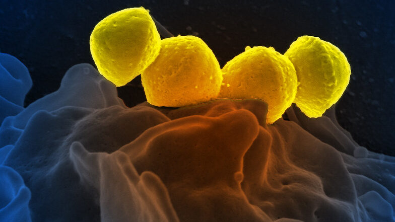
Streptococcus pyogenese bound to a human neutrophil. Credit: National Institute of Allergy and Infectious Diseases, National Institutes of Health
Bordering on science fiction, medicinal microrobots could help physicians better treat and prevent diseases. But a serious problem is the synthetic materials they are made of trigger immune responses. Now, for the first time, researchers report in ACS Central Science that they achieved precise control neutrophils as a natural, biocompatible microrobot by using lasers. By getting the ‘neutrobots’ to perform multiple tasks, the researchers demonstrated they could one day deliver drugs to precise locations in the body.
Microrobots being developed for medical applications would need to be administered in injections or oral capsules to get them inside the body. But these microscopic objects are often found to trigger immune reactions in small animals, resulting in the the microrobots being ejected from the body before they can carry out their tasks. By using the body’s own cells, such as neutrophils, drugs could be delivered less invasively without provoking an immune response.
Neutrophils already naturally pick up nanoparticles and dead red blood cells and can migrate through blood vessels into adjacent tissues, so they are good candidates for becoming microrobots. Previously, researchers have guided neutrophils with lasers in lab dishes, moving them around as ‘neutrobots’. However, this had not been tried in living animals. So, researchers set out to demonstrate the feasibility of light-driven neutrobots in animals using live zebrafish.
The researchers manipulated and moved neutrophils in zebrafish tails, using focused laser beams as optical tweezers. The ‘neutrobots’ could be moved up to a velocity of 1.3 µm/s, three times faster than a neutrophil’s natural speed. The optical tweezers were able to precisely and actively control the functions that neutrophils conduct as part of the immune system. For example, moving through a blood vessel wall into the surrounding tissue; carrying a plastic nanoparticle, showing potential for delivering medicine; and pushed towards red blood cell debris, a neutrophil engulfed the pieces. Surprisingly, at the same time, a different neutrophil, which wasn’t controlled by a laser, tried to naturally remove the cellular debris. Because they successfully controlled neutrobots in vivo, the researchers say this study advances the possibilities for targeted drug delivery and precise treatment of diseases.
About a third of people with psoriasis develop inflammation in their joints (psoriatic arthritis) as a result of the chronic skin condition. Research published in Annals of the Rheumatic Diseases has now discovered a key starting point for inhibiting inflammation in both psoriasis and psoriatic arthritis. These findings may lead to major new developments for treatment, diagnostic and prevention strategies.
The study conducted by the research group led by Erwin Wagner at the Medical University of Vienna focused on the S100A9 gene. The team has discovered that the severity of psoriasis (Ps) and psoriatic arthritis (PsA) can be reduced by inhibiting S100A9 systemically throughout the whole body rather than locally on the skin.
With this finding, the researchers are laying the foundation for a paradigm shift in the treatment of Ps and PsA: “Our study is an important step towards the development of targeted therapeutic options in the form of drugs that act systemically rather than locally on the skin,” affirms Erwin Wagner. New diagnostic and prevention strategies can also build on the study.
Psoriasis, typically an adult-onset disease, have triggers such as stress and UV radiation. There can also be a genetic predisposition to developing Ps. S100A9 activation in skin and immune cells has been identified as a risk factor for the development of Ps and/or PsA.
Previous work by Erwin Wagner’s team showed that the symptoms of psoriasis disappear when the S100A9 gene is deactivated in all of the body’s cells. Their recent preclinical experiments highlighted the particular influence that those skin and immune cells in which S100A9 is produced have on disease severity. “We now know that the inflammatory responses in psoriasis and psoriatic arthritis are enhanced when S100A9 is only inhibited in skin cells,” Erwin Wagner explained. Therefore drugs inhibiting S100A9 would have to be administered systemically in the form of tablets or drips
In 2016, Spinraza® became the by the first FDA-approved treatment for spinal muscular atrophy (SMA). This neurodegenerative disease is the leading genetic cause of infant death. The drug was conceived and developed by Cold Spring Harbor Laboratory (CSHL) Professor Adrian Krainer and collaborators. But Prof Krainer’s lab continued to try and improve Spinraza® could be improved, in collaboration with Alberto Kornblihtt at Universidad de Buenos Aires. They discovered pairing Spinraza® with a second FDA-approved drug called valproic acid (VPA) could be a new way to boost its therapeutic effects, without increasing toxic side effects.
Prof Krainer explained: “Sometimes you don’t want to use a ton of a drug. If you have a condition that allows you to use less drug, then you may have fewer toxicities. So the idea is to combine these two drugs to get maximal effects.”
In SMA, the body produces insufficient amounts of a protein called SMN. Spinraza® is a type of molecule called an antisense oligonucleotide (ASO) that helps cells make more SMN protein from a gene called SMN2. Roadblocks were discovered on the SMN2 gene when using Spinraza®, slowing down the cellular machine producing SMN protein. The drug VPA helps remove these roadblocks, allowing Spinraza® to further increase the SMN protein output. When mice with SMA were treated with both VPA and a Spinraza®-like ASO used for research, the mice survived longer, with improved muscle function.
To date, more than 11 000 SMA patients have been treated with Spinraza® in more than 50 countries. Prof Krainer’s latest research shows that there’s always room for improvement. He hopes the team’s findings will help optimize the efficacy of Spinraza® treatments, and hopes their work will help the development of treatments for other neurodegenerative diseases.
Healthcare workers are among the 20% of the world’s population who do shift work. Shift workers’ differing sleep-wake cycle is a risk factor for numerous health disorders, including diabetes, heart attacks, cancer and strokes.
Now, new research published in Neurobiology of Sleep and Circadian Rhythms shows the adverse effects of shift work can be long-lasting, even after returning to a normal schedule.
“Shift work, especially rotating shift work, confuses our body clocks and that has important ramifications in terms of our health and well-being and connection to human disease,” said Professor David Earnest at the Texas A&M University College of Medicine. “When our internal body clocks are synchronized properly, they coordinate all our biological processes to occur at the right time of day or night. When our body clocks are misaligned, whether through shift work or other disruptions, that provides for changes in physiology, biochemical processes and various behaviours.”
A previous study done by Earnest and colleagues found that animal models on rotating shift work schedules had more severe stroke outcomes, than those on regular 24-hour cycles of day and night. Males were distinguished by worse outcomes in which mortality rates were much higher.
This new study took a different approach. Rather than examining immediate effects of shift work on strokes, the researchers returned all subjects to regular 24-hour cycles and waited until their midlife equivalent – when humans are most likely to experience a stroke – to evaluate stroke severity and outcomes.
“What was already born out in epidemiological studies is that most people only experience shift work for five to eight years and then presumably go back to normal work schedules,” Prof Earnest said. “We wanted to determine, is that enough to erase any problems that these circadian rhythm disruptions have, or do these effects carry over even after returning to normal work schedules?”
They found that the health impacts of shift work do, indeed, persist over time. The sleep-wake cycles of subjects on shift work schedules never truly returned to normal, even after subsequent exposure to a regular schedule. Compared to controls maintained on a regular day-night cycle throughout the study, they displayed persistent alterations of their sleep-wake rhythms, with periods of abnormal activity when sleep would have normally occurred. When they suffered strokes, their outcomes were again much worse than the control group, except females had more severe functional deficits and higher mortality than the males.
“The data from this study take on added health-related significance, especially in females, because stroke is a risk factor for dementia and disproportionately affects older women,” said Professor Farida Sohrabji.
The researchers also observed increased levels of inflammatory mediators from the gut in subjects exposed to a shift work schedule. “We now think that part of the underlying mechanism for what we’re seeing in terms of circadian rhythm disruption causing more severe strokes may involve altered interactions between the brain and gut,” Prof Earnest said.
The results of this study could eventually lead to the development of interventions that block adverse effects of disrupted circadian rhythms. In the meantime, shift workers can improve care of their internal body clocks by trying to maintain a regular schedule as much as possible and avoiding a high-fat diets, which can cause inflammation and also alter the timing of circadian rhythms.
This research has clear implications for shift workers, but it could extend to many other people who keep schedules that differ greatly from day to day. Modern work has also extended the work day thanks to email and the internt.
“Because of the computer age, many more of us are no longer working from nine to five. We take our work home and sometimes work late at night,” Prof Earnest said. “Even those of us who do work regular schedules have a tendency to stay up late on the weekends, producing what is known as ‘social jet lag,’ which similarly unwinds our body clocks so they no longer keep accurate time. All this can lead to the same effects on human health as shift work.”
Research into the mechanisms of cell migration and the impact of tissue rigidity on cell positioning and steering has found that cancer cell migrate towards and opens new possibilities for stopping and directing it.
An international team of scientist have uncovered for the first time how tissue stiffness determines cell positioning and regulates all types of cell migration ranging from the neuronal growth cone turning to dissemination of malignant cancer cells in brain tumors and breast cancer.
Each cell in the body has a specific task and carefully determined position within a tissue. Cell positioning is regulated by many factors, including tissue rigidity. Cells are capable of probing and sensing their environment, and different cell types have different preferences for optimal conditions. A little bit like Goldilocks in the story trying out the different beds of the bear family and finding one bed to be too soft, the other one two hard and one to be just right. While this has been well-known for a long time, it has remained a mystery to researchers how cells are able to steer themselves to the optimal environment.
“The prevailing view among scientists was that all cell types prefer high-rigidity environments and migrate towards increasing stiffness. This process has been coined the term ‘durotaxis‘ – migration towards hard from Greek and Latin,” said Aleksi Isomursu, a doctoral researcher.
“I was visiting the University of Minnesota for a research project and noticed that brain cancer cells grown on engineered substrates with alternating stiffness show the opposite behaviour they turned towards soft,” Isomursu continues.
This observation launched an interdisciplinary research project involving cancer cell biology, computational modelling and engineering and involving researchers from three continents. As the outcome, the researchers uncovered the basic mechanism all cell types use to steer themselves towards their optimal environment.
These results will be important for future research in stopping and directing cancer cell migration.
“I experimented with different types of drugs and identified ones that could make brain cancer cells stop moving or change direction,” explains Postdoctoral Researcher Mathilde Mathieu.
Identification of the mechanism of cell steering provides explanations for many thus far mysterious steps in cancer dissemination, for example how cancer cells migrate out from the stiff core of a breast tumour.
“These findings have been gaining a lot of interest in researchers and we have even played around with the idea of launching a new term – ‘mollitaxis‘, migration towards soft,” says the Principal Investigator of the laboratory at the University of Turku, Professor Johanna Ivaska.
Patients identified by nuclear stress testing as having severe stress-induced myocardial ischaemia may benefit from angioplasty, while those with mild or no ischaemia will not, according to a new study reported in the Journal of the American College of Cardiology.
Following stress testing, coronary revascularisation restores blood flow to blocked arteries. For patients with severe ischaemia, early revascularisation saw a more than 30% reduction in mortality compared to patients with severe ischaemia who were treated with medication, but there was no benefit for the other groups.
Conducted by the Icahn School of Medicine at Mount Sinai, this is the first large-scale study to investigate stress testing in patient management when applied to the full spectrum of patients who have both varying degrees of myocardial ischaemia and heart function. This new study can help guide physicians on how to manage caring for patients with suspected heart disease.
Stress tests are indicated when physicians suspect that a patient’s chest pain or other clinical symptoms are from coronary artery disease (CAD). These help determine if a patient has obstructive CAD which leads to significant ischaemia. If the ischaemia due to obstructive CAD is severe, adequate blood flow can be restored with coronary artery bypass grafting surgery or percutaneous coronary intervention (PCI), where a catheter is used to place stents in the blocked coronary arteries. Nuclear stress testing is the most common stress test used to detect myocardial ischaemia.
“There is keen interest in assessing how measurement of myocardial ischaemia during stress testing can help shape physicians’ decision to refer patients for coronary revascularisation procedures, but this issue has not been well studied among patients who have underlying heart damage,” explains lead author Alan Rozanski, MD. “Our study, which evaluated a large number of patients with pre-existing heart damage who underwent cardiac stress testing, finally addresses this clinical void.”
The researchers analysed records of more than 43 000 patients who underwent nuclear stress testing with suspected CAD between 1998 and 2017 with a median 11-year follow-up for mortality/survival. The investigators grouped patients according to both their level of myocardial ischaemia during stress testing as well as their left ventricular ejection fraction (LVEF). Low LVEF measurements indicate prior heart damage that could be from scarring of the heart due to a prior heart attack.
The study provides two important clinical insights. First, the study showed that the frequency of myocardial ischemia during stress testing varies according to patients’ heart function. Of the 39 883 patients with normal heart function (LVEF > 55%), fewer than 8% of them had ischaemia. However, among the 3560 patients with reduced heart function (LVEF less than 45%, which indicates prior heart damage), more than 40% of them had myocardial ischaemia. The study also showed that the presence of myocardial ischaemia increases the risk of death in patients with normal and reduced heart function. Among both groups of patients, performing bypass or PCI procedures was not associated with improved survival among patients with either no or only mild ischaemia during the cardiac stress test. Among patients with severe ischaemia, coronary procedures were associated with more than 30% higher survival rates compared to those managed with medication only. This was the case for patients with and without heart damage.
“These results confirm the benefits of stress testing for clinical management. What you want from any test when considering coronary revascularisation procedures is that the test will identify a large percentage of patients who are at low clinical risk and do so correctly, while identifying only a small percentage of patients who are at high clinical risk and do so correctly. That is what we found with nuclear stress testing in this study,” explains Dr Rozanski. “Importantly, the presence of severe ischaemia does not necessarily mean that coronary revascularisation should be applied. New data from a large clinical trial suggests that when medical therapy is optimised it may be as effective as coronary revascularisation in such patients. But regardless, the presence of severe ischaemia indicates high clinical risk which then requires aggressive management to reduce clinical risk.”
A candidate cancer drug currently in development has been also shown to stimulate regeneration of damaged nerves after spinal trauma. Four weeks after spinal cord injury, animals treated with the candidate drug, AZD1390, were “indistinguishable” from uninjured animals, according to the researchers.
The study, published in Clinical and Translational Medicine, demonstrated in cell and animal models that the candidate drug, AZD1390, can block the response to DNA damage in nerve cells and promote regeneration of damaged nerves. This restored sensory and motor function after spinal injury.
The announcement comes weeks after the same research team showed a different investigational drug, AZD1236, can reduce damage after spinal cord injury, by blocking the inflammatory response.
AZD1390 is also under investigation by AstraZeneca to block ATM-dependent signalling and repair of DNA double strand breaks (DSBs), an action which sensitises cancer cells to radiation treatment. The ATM protein kinase pathway – a critical biochemical pathway regulating the response to DNA damage. The DNA Damage Response system (DDR) is activated by DNA damage, including DSBs in the genome, which also happen in several common cancers and also after spinal cord injury.
Professor Zubair Ahmed, from the University’s Institute of Inflammation and Ageing and Dr Richard Tuxworth from the Institute of Cancer and Genomic Sciences hypothesised the persistent activation of this system may prevent recovery from spinal cord injury, and that blocking it would promote nerve repair and restore function after injury.
Their initial studies found that AZD1390 stimulated nerve cell growth in culture, and inhibited the ATM protein kinase pathway – a critical biochemical pathway regulating the response to DNA damage.
AZD1390 was tested in animal models following spinal cord injury. Oral treatment with AZD1390 significantly suppressed the ATM protein kinase pathway, stimulated nerve regeneration beyond the site of injury, and improved the ability of these nerves to carry electrical signals across the injury site.
Professor Ahmed commented: “This is an exciting time in spinal cord injury research with several different investigational drugs being identified as potential therapies for spinal cord injury. We are particularly excited about AZD1390 which can be taken orally and reaches the site of injury in sufficient quantities to promote nerve regeneration and restore lost function.
“Our findings show a remarkable recovery of sensory and motor functions, and AZD1390-treated animals being indistinguishable from uninjured animals within 4 weeks of injury.”
Dr Tuxworth added: “This early study shows that AZD1390 could be used as a therapy in life-changing conditions. In addition, repurposing this existing investigational drug potentially means we can reach the clinic significantly faster than developing a new drug from scratch.”
p53 is one of the most important proteins in cancer biology. Often referred to as a “guardian of the genome”, p53 activates in response to cellular stressors such as DNA damage. Its activation induces processes such as cell death that prevent cancer development. Because of this, p53 mutations are extremely common in cancers, including hepatocellular carcinoma.
However, a study published in Cancer Research, revealed that the constant activation of p53 in liver cells of patients suffering from chronic liver disease (CLD) could actually promote the development of liver cancer.
CLD can be brought on by different factors including viruses, alcohol use, and fat accumulation, all of which can induce p53 activation. Previous studies have shown that p53 is in a constant state of activation in the liver cells of CLD patients. Yet, it is not clear what role this plays in CLD pathophysiology.
“Clinical data clearly show that p53 is activated in the hepatocytes of individuals with CLD,” says Yuki Makino, lead author of the study at Osaka University. “Because p53 is such a vital part of how the human body prevents tumor formation, its role in CLD became even more intriguing.”
To create a mouse model with p53 accumulation in hepatocytes, researchers deleted Mdm2, the protein responsible for regulating p53 expression by targeting it for degradation. These mice developed liver inflammation with higher amounts of hepatocyte apoptosis and senescence-associated secretory phenotype (SASP), a phenomenon where cells produce signals within the microenvironment that can cause nearby cells to become cancerous. In fact, mice with p53 accumulation did have increased liver tumour development.
“We also observed an expanded population of hepatic progenitor cells (HPCs), which have stem cell-like characteristics,” explained senior author Tetsuo Takehara. “When the HPCs were isolated, grown in culture, and then injected under the skin of lab mice, these animals developed tumors. This suggested that HPCs played a key part in the liver tumor formation seen in the animals with p53 accumulation.”
Interestingly, acceleration of liver tumour development and the other observed phenotypes did not occur when p53 was deleted in addition to Mdm2 in the hepatocytes. These results demonstrated the significance of constant p53 activity in the tumorigenesis.
“We then compared samples from 182 CLD patients with 23 healthy liver samples,” said Dr Makino. “The CLD liver biopsy samples showed activated p53 was positively correlated with apoptosis levels, SASP, HPC-associated gene expression, and later cancer development.”
The authors concluded that constitutively activated p53 in hepatocytes of CLD patients can create a microenvironment that is supportive of tumour formation from HPCs. Their work proposes a novel and paradoxical mechanism of liver tumorigenesis because p53 is one of the most well-known tumor suppressor genes. These data could highlight p53 as a potential cancer-prevention treatment target for CLD patients.