Acute low back pain is a common cause of disability, and while opioid drugs are effective at controlling pain, excessive use creates a great potential for substance abuse. An analysis in the Journal of Orthopaedic Research examined which non-opioid drugs are best for relieving this pain.
The analysis, which included all randomised controlled trials published to date (18 studies with 3478 patients), showed that muscle relaxants and non-steroidal anti-inflammatory drugs (NSAIDs) could effectively and rapidly reduce symptoms.
The combination of NSAIDs and paracetamol was associated with a greater improvement than NSAIDs alone.
“This is a first step towards the optimisation of the management of acute low back pain. However, specific patient characteristics such as having allergies and comorbidities must always be taken into consideration,” said lead author Alice Baroncini, MD, PhD, of RWTH University Hospital in Germany. “Further research will need to focus on the identification of the type of drugs that not only offer the best and quickest pain relief, but also show the lowest rate of symptom recurrence.”
A study published in PLoS ONE has confirmed the role of the corpus callosum in language lateralisation, ie the distribution of language processing functions between the brain’s hemispheres. To get to this finding, the researchers applied advanced neuroimaging methods to study subjects performing an innovative language task for their study.
Functional asymmetry between the two cerebral hemispheres in performing higher-level cognitive functions is a major characteristic of the human brain. For example, the left hemisphere plays a leading role in language processing in most people. However, between 10% and 15% of people also use the right hemisphere to varying degrees for the same task.
Traditionally, language lateralisation to the right hemisphere was explained by handedness, as it is mainly found in left-handed and ambidextrous (using both hands equally well) individuals. But recent research has demonstrated a genetic difference in the way language is processed by left-handed and ambidextrous people. In addition to this, some right-handed people also involve their right hemisphere in language functions.
These findings prompted the scientists to consider alternative explanations, especially by looking at brain anatomy to find out why language functions can shift to the right hemisphere. Researchers at the HSE Centre for Language and Brain hypothesised that language lateralisation may have something to do with the anatomy of the corpus callosum, the largest commissural tract in the human brain connecting the two cerebral hemispheres.
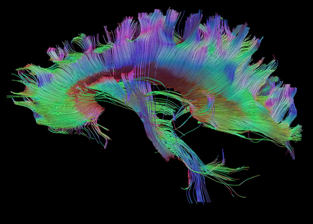
Tractography of the corpus callosumDo Tromp et al. / brainimaging.waisman.wisc.edu
The researchers asked 50 study participants to perform a sentence completion task. The subjects were instructed to read aloud a visually presented Russian sentence and to complete it with an appropriate final word (eg ‘Teper’ ministr podpisyvaet vazhnoe…‘ – ‘Now the minister is signing an important …’). At the same time, the participants’ brain activity was recorded using functional magnetic resonance imaging (fMRI). Additionally, the volume of the corpus callosum was measured in each subject.
A comparison between the fMRI data and the corpus callosum measurements revealed that the larger the latter’s volume, the less lateralisation of the language function to the right hemisphere was observed.
When processing language, the brain tends to use the left hemisphere’s resources efficiently and the corpus callosum suppresses any additional involvement of the right hemisphere. The larger a person’s corpus callosum, the less involved their right hemisphere is in language processing (and vice versa). This finding is consistent with the inhibitory model suggesting that the corpus callosum inhibits the action of one hemisphere while the other is engaged in cognitive tasks.
The study’s innovative design and use of advanced neuroimaging have made this conclusion possible. Brain lateralisation in language processing is usually hard to measure accurately, as typical speech tasks used in earlier studies (eg image naming, selecting words that begin with a certain letter or listening to speech) tend to cause activation only in some parts of the brain responsible for language functions but not in others. Instead, we developed a unique speech task for fMRI: sentence completion, which reliably activates all language areas of the brain.
The researchers reconstructed the volume and properties of the corpus callosum from MRI data using an advanced tractography technique: constrained spherical deconvolution (CSD). This is more suitable than traditional diffusion tensor imaging for modelling crossing fibres in the smallest unit of volume, the voxel (3D pixel), and is therefore more reliable.
A new poll done in the US suggests that some parents may not be properly measuring or responding to elevated temperatures in children, and are unnecessarily using antipyretics to bring down their temperatures.
While most parents recognise that a low-grade fever helps a child’s body fight off infection, one in three would give fever-reducing medication for spiked temperatures below 38°C (which isn’t recommended) according to the C.S. Mott Children’s Hospital National Poll on Children’s Health at University of Michigan Health.
Half of parents would also use medicine if the fever was between 38 and 38.9°C, and a quarter of parents would likely give another dose to prevent the fever from returning.
“Often parents worry about their child having a fever and want to do all they can to reduce their temperature. However, they may not be aware that in general the main reason to treat a fever is just to keep their child comfortable,” said Mott Poll co-director and Mott pediatrician Susan Woolford, M.D.
“Some parents may immediately rush to give their kids medicine but it’s often better to let the fever runs its course. Lowering a child’s temperature doesn’t typically help cure their illness any faster. In fact, a low-grade fever helps fight off the infection. There’s also the risk of giving too much medication when it’s not needed, which can have side effects.”
The report is based on 1,376 responses from parents of children ages 12 and under polled between August and September 2022.
Two in three parents polled say they’re very confident they know whether their child needs medication to reduce a fever. But just over half are sure they understand how temperature readings can change according to the method used.
The method used to take a child’s temperature matters and can affect the accuracy of the measurement, Woolford notes. Parents polled most commonly take their child’s temperature by forehead scan or mouth while less than a sixth use ear, underarm or rectal methods.
Remote thermometers at the forehead or inside the ear canal can be accurate if used correctly. But forehead readings may be inaccurate, Woolford says, if the scanner is held too far away or if the child’s forehead is sweaty. With ear thermometers, which aren’t recommended for newborns, earwax can also interfere with the reading.
For infants and young children, rectal temperatures are most accurate. Once children are able to hold a thermometer in their closed mouth, oral temperatures also are accurate while armpit temperatures are the least accurate method.
“Contact thermometers use electronic heat sensors to record body temperature but temperatures may fluctuate depending on how it’s measured,” Woolford said.
“Regardless of the device used, it’s important that parents review the directions to ensure the method is appropriate for the child’s age and that the device is placed correctly when measuring temperature.”
Three in four parents say they take their child’s temperature as soon as they notice a possible problem, while a little less than a fourth wait to see if the problem continues or worsens before taking the temperature.
Two-thirds of parents also prefer to try methods like a cool washcloth before using fever-reducing medication. Most parents also say they always or usually record the time of each dose and re-take their child’s temperature before giving another dose.
“A quarter of parents would give their child more medicine to prevent a fever from returning even though it doesn’t help them get better,” Woolford said. “If a child is otherwise doing well, parents may consider monitoring them and using alternative interventions to help keep them comfortable.”
However, if a newborn or infant less than three months old has a fever, they should immediately see a health professional, Woolford adds.
Researchers have found that compounds in the edible Lions’ Mane mushroom (Hericium erinaceus), already used in herbal medicine for stomach complaints, can promote nerve growth and boost memory. Their findings are published in The Journal of Neurochemistry.
The compounds are neurotrophins – a family of proteins associated with the growth, functioning and maintenance of neurons. In mammals, brain-derived neurotrophic factor (BDNF) is highly expressed in central nervous system neurons. Because BDNF pathway impairment is associated with several diseases, including schizophrenia, Alzheimer’s disease, Rett syndrome, and Huntington’s disease, experimental treatments for neurological and neurodegenerative disorders often target neutrophins.
So far, such treatments have run into problems with crossing the blood-brain barrier and off-target effects. H. erinaceus is high in neutrophins, though it has traditionally been used in herbal medicine to treat stomach complaints and cancer. It is also known for promoting peripheral nerve regeneration.
Two of the biologically active types of compounds, hericenones and erinacines, can successfully cross the the blood-brain barrier and confer neuroprotective effects.
The researchers tested crude and purified H. erinaceus extracts and found that they exhibited BDNF-like neurotrophic activity in both in vitro cultured hippocampal neurons and in mouse models of hippocampal memory. The extracts also promoted neurite outgrowth and improved memory.
They concluded that Hericene A acts through a novel signalling pathway, giving rise to improved cognitive performance. These findings however will need to be validated by future research.
Researchers have discovered that the difference between people who do and do not respond to immunotherapy may have to do with an immune cell known as CD5+ dendritic cells, because they bear the protein CD5 on their outer surfaces. Their research, published in Science, showed that survival was longer in patients with a range of cancers, including melanoma, if they had more CD5+ dendritic cells in their tumours, and that mice lacking CD5 on their dendritic cells failed to respond well to immunotherapy.
The findings suggest that a supplementary therapy designed to increase the number or activity of CD5+ dendritic cells potentially could extend the lifesaving benefits of immunotherapy to more cancer patients.
“Immunotherapy has revolutionized the field of cancer therapy, but there are a lot of patients with cancer who don’t benefit from it,” said senior author Eynav Klechevsky, PhD, an assistant professor of pathology & immunology and a researcher at Siteman Cancer Center at Barnes-Jewish Hospital and Washington University School of Medicine. “Part of the reason some people do not respond well to some forms of immunotherapy is because this population of dendritic cells is reduced dramatically. We’re developing some novel immune-based approaches to boost the activation of these CD5-expressing dendritic cells with a goal of helping more patients respond to immunotherapy.”
The immune system defends the body against cancer by activating immune cells known as T cells to recognise and kill tumour cells. In response, tumour cells manipulate the immune checkpoint system — a safeguard that prevents T cells from mistakenly attacking healthy cells — to hoodwink T cells into leaving them alone. Immune checkpoint blockade therapy works by thwarting tumour cells’ manipulations, thereby freeing T cells to recognise and destroy tumours. But even with therapy, some people’s T cells are unable to do their job effectively.
Klechevsky and colleagues — including first author Mingyu He, PhD, a staff scientist, and co-author Kate Roussak, MD, a postdoctoral researcher — suspected that people who don’t respond to immunotherapy may have a problem with their dendritic cells. Without dendritic cells, T cells are subdued and aimless.
By analysing data in The Cancer Genome Atlas, Klechevsky and colleagues discovered that patients with types of skin, lung, bone and soft tissue, breast and cervical cancers fared better if they had higher levels of CD5+ dendritic cells in their tumours.
Further work with human cells and mice showed that CD5+ dendritic cells are required for effective T cell activity against tumours. CD5+ dendritic cells from people powerfully induced T cells to activate and multiply. Mice with tumours responded only weakly to immunotherapy and failed to reject the tumors if they lacked CD5 on their dendritic cells.
The findings suggest that the amount of CD5+ dendritic cells inside tumours could be used to help doctors assess which patients are most likely to benefit from immunotherapy. They also suggest that increasing the numbers or the activity of such dendritic cells potentially could help more people benefit from immunotherapy. As part of this study, the researchers discovered that the immune protein IL-6 increases the amounts of CD5+ dendritic cells.
“We still don’t completely understand how immunotherapies work,” Klechevsky said. “This study indicates that there is more we can do to increase the efficacy of these treatments. I’m confident that if we can find ways to harness these cells or expand these cells in patients, we can help more people.”
Changing the structure of a dietary fibre commonly found in a range of food products has been found to promote healthy gut bacteria and reduce gas formation, a finding that could help people with intolerances to fibre and irritable bowel conditions. The findings have been published in Food Hydrocolloids.
A team of scientists from the University of Nottingham, Quadram Institute Biosciences and the University of East Anglia examined psyllium, a type of natural dietary fibre that is used in a range of products including cereals and yoghurts. They showed that the physical state of the fibre has a major impact on gas production which often is linked to bowel discomfort.
The team performed in vitro fermentation experiments seeded with human stool. They conducted analysis of fermentation products and evaluated the impact of different structures on the broad categories of microorganisms.
Dr Gleb Yakubov, Associate Professor in Food Physics at the University of Nottingham was one of the lead researchers on the study, he explains: “Although fibre is an important part of any diet, for many people it can cause bowel discomfort and for people with IBS or IBD fibre can be a trigger. This is because some foods cause bacterial interactions in the gut that create gas that can lead to pain or discomfort. Our study shows that the physical state of the fibre has a major impact on gas production by creating beneficial compounds that promote the creation of the good bacteria in the gut.”
Psyllium fibre comes from the seeds of Plantago ovata plants, known by many common names such as blond plantain. These seeds produce a jelly-like material called mucilage, which comes in a variety of shapes and forms and these feature long-chain sugars, called polysaccharides. It is these polysaccharides that lead to the production of beneficial short-chain fatty acids that contribute positively to gut health and systemic metabolism. This study shows that different physical states of fibre impact the way dietary fibre breaks down and that microbes ‘colonise fibre’ during fermentation.
Professor Yakubov continues: “These findings show that there are new opportunities for designing targeted structures using psyllium, either through seed processing or selective breeding, to achieve new fibre materials with clear clinical benefit above that of unrefined psyllium powders aiding in the treatment of gastrointestinal discomfort.”
Research is already underway to create and test psyllium-mimicking materials as medical nutrition which could provide a source of fibre for people with some bowel conditions.
A new study published in Science Advances shows that female and male hearts respond differently to the stress hormone noradrenaline. The study in mice may have implications for human heart disorders like arrhythmias and heart failure and how different sexes respond to various drugs.
Using fluorescence imaging, the researchers were able to see in real time and in vivo how a mouse heart responds to hormones and neurotransmitters, including noradrenaline.
The results reveal that male and female mouse hearts respond uniformly at first after exposure to noradrenaline. However, some areas of the female heart return to normal more quickly than the male heart, producing differences in the heart’s electrical activity.
“The differences in electrical activity that we observed are called repolarisation in the female hearts. Repolarisation refers to how the heart resets between each heartbeat and is closely linked to some types of arrhythmias,” said Jessica L. Caldwell, first author of the study.
“We know that there are sex differences in the risk for certain types of arrhythmias. The study reveals a new factor that may contribute to different arrhythmia susceptibility between men and women,” Caldwell said.
Methods
The novel imaging system uses a genetically modified ‘CAMPER’ mouse to emit light during a very specific chemical reaction in the heart: cAMP binding.
The cAMP molecule (an abbreviation of cyclic adenosine 3′,5;-monophosphate) is an intermediate messenger that turns signals from hormones and neurotransmitters, including noradrenaline, into action from heart cells.
The light signals from the CAMPER mouse are transmitted by a biosensor that uses a fluorescence signal that can be picked up at high speed and high resolution by a new imaging system specially designed for hearts. This allows the researchers to record the heart’s reaction to noradrenaline in real time, along with changes in electrical activity.
This new imaging approach revealed the differences in the breakdown of cAMP in female and male mice and the associated differences in electrical activity.
Including female mice leads to discoveries
The researchers had not planned to study sex-based responses, according to Crystal M. Ripplinger, senior author of the study. But the researchers started seeing a pattern of different reactions, which led them to realise the differences were sex-based.
When Ripplinger started her lab at the UC Davis School of Medicine over a decade ago, she exclusively used male animals. That was the norm for most research at the time. But several years ago, she began including male and female animals in her studies.
“Sometimes the data between the two sexes is the same. But if the data start to show variation, the first thing we do is look at sex differences. Using both male and female mice has revealed clues into differences we would never have suspected. Researchers are realising you can’t extrapolate to both sexes from only studying one,” Ripplinger said.
She notes that with the current study, it’s not clear what the differences in cAMP and electrical activity may mean.
“The response in the female mice may be protective – or it may not. But simply documenting that there is a measurable difference in the response to a stress hormone is significant. We are hoping to learn more in future studies,” Ripplinger said.
One early indicator of Parkinson’s disease (PD) is isolated REM-sleep behaviour disorder. Researchers have shown that a greater concentration of α-synuclein aggregates can be detected in the stool samples of patients. In the scientific journal npj Parkinson’s Disease, they now present a method for detecting these aggregates.
There are two forms of PD. In 70% of cases, it originates in the central nervous system. However, in around 30% of cases it originates in the nervous system of the intestine (“enteric nervous system”). The latter form is referred to as “body-first Parkinson’s disease” (for short: body-first PD) and the characteristic deposits of aggregates of the body’s own α-synuclein protein are formed in the neurons in the intestine.
A preliminary form of body-first PD is the so-called isolated REM-sleep behaviour disorder (for short: iBRD). It causes in part complex movements during REM-sleep insofar as the patient experiences vivid and disturbing dreams. These movements can endanger the sufferer themselves or others.
A research team headed by Professor Erdem Gültekin Tamgüney from the Institute of Physical Biology at HHU now reports that it is possible to detect an elevated level of α-synuclein aggregates in the stool samples of patients. To achieve this, the team used a new surface-based fluorescence intensity distribution analysis (sFIDA) to detect and quantify individual particles of α-synuclein aggregates.
Professor Tamgüney: “We are the first to prove the presence of α-synuclein aggregates in stool samples. Our results show a significantly higher level of α-synuclein aggregates in iRBD patients compared with healthy individuals or patients with Parkinson’s. These findings could lead to a non-invasive diagnostic tool for prodromal synucleinopathies — including Parkinson’s — which could in turn enable therapies to be initiated at an early stage before symptoms occur.” However, more research is required before the process can find its way into clinical practice, for example investigation into why the level is lower in Parkinson’s patients.
The study was conducted in a collaboration to establish a biobank with stool samples from patients and control subjects, and to develop the test procedure and conduct the tests on the samples, and to eventually commercialise the technique.
Background
In body-first PD, the deposits of fibrils of the body’s own α-synuclein protein, which are characteristic of Parkinson’s, are first formed in the neurons of the enteric nervous system, which serves the gastrointestinal tract. The aggregates then spread to the central nervous system in a way similar to prions, i.e. an existing aggregate combines individual α-synuclein proteins in its vicinity into further aggregates in a nucleation process; these aggregates then spread further through the body.
The influence of what happens in the gastrointestinal tract on the brain is referred to as the “gut-brain axis.” The gastrointestinal tract is exposed to the environment and it is possible that harmful substances such as chemicals, bacteria or viruses ingested directly with food or via interaction with the microbiome of the gastrointestinal tract may trigger the pathological formation of α-synuclein aggregates.
A Bloomberg investigation has revealed that Glaxo, which went on to form pharmaceutical giant GSK, was aware of the possible carcinogenic content of its popular heartburn drug Zantac for over 40 years.
The once-popular heartburn drug Zantac (ranitidine) was developed in the 1970s by the then-small British pharmaceutical company Glaxo. It was marketed as better and safer than Tagamet (cimetidine) and it soon outsold it and became one of the company’s best-selling drugs.
NDMA belongs to a class of compounds known as nitrosamines, came to light in cancer research when it was found that nitrites could combine with amines in the stomach, giving rise to cancer-causing compounds. This led to calls for restrictions of sodium nitrites, found in various cured foods, but the food industry has resisted this.
This all came to an end in 2019, when Zantac was found to be contaminated by the chemical N-Nitrosodimethylamine (NDMA), a pale yellow liquid at room temperature. NDMA is a known carcinogen at high concentrations and a possible one at low concentrations. This contamination was not due to a manufacturing error, however: NDMA can form from other reactions and in this case came from within the drug itself. Around the world, Zantac was pulled from shelves, and in 2020 the drug was banned by the FDA. NDMA currently is only produced in small amounts to induce cancer in rats. Whether it is carcinogenic at very low levels is still a matter of debate, but Zantac products contained NDMA at levels which could increase over time.
According to Bloomberg, Glaxo was aware of this, as the possibility of ranitidine being converted in the stomach to nitrosamines being raised in 1980. Tests with human subjects taking ranitidine showed this. Even though NDMA is widely held to be a carcinogen in humans, it is difficult to prove that a particular chemical is responsible for mutations leading to cancers. Glaxo simply took the view that ranitidine did not cause cancer in rodent studies, and that any possible cancer risk was minimised by short exposure. In fact, users would take Zantac for extended periods, even years or decades.
The company was thus warned over 40 years by its own scientists as well as independent researchers, with thousands of pages of documentation seen by Bloomberg for the first time. One 1981 Lancet study showed that Zantac mixed with nitrite in the stomach (as from a meal) would produce nitrosamines. Glaxo scientists countered by saying that the nitrite levels in the study were far above those found in a human stomach, a defence which would become their standard response.
While Glaxo was preparing for FDA approvals in 1982, another study came to their attention. Concerned, Glaxo asked one of their scientists, Richard Tanner, to replicate it and he found that some samples contained 232 000ng of NDMA – the FDA approved limit for each drug was 96ng, though with lower nitrite levels, no NDMA was detected. The daily FDA exposure limit from all sources for NDMA is 190ng, which includes NDMA from all sources, including other medications (which may produce their own NDMA over time) as well as biological processes.
This report would remain secret for more than 40 years. More trouble came from the fact that NDMA could be formed from exposure to either heat or humidity. When it went through the FDA approval process, it did not mention the Tanner report and the notion of storage was glossed over: approved in 150mg doses for eight weeks, storage requirements were listed as a dry place at a temperature no higher than 86°F (30°C).
Zantac surged in popularity, turning the company into a multi-billion dollar enterprise, though the FDA took action over Glaxo’s claims. An over-the-counter 75mg version was launched in 1996. It was coloured pink due to issues of discolouration with white pills turning yellow or brown over time.
While discolouration issues were briefly investigated, it was only in 2019 when there were concerns over NDMA contamination, spurred by recalls of a blood pressure drug, valsartan. A private laboratory warned the FDA over NDMA discovery in Zantac and ranitidine generics, and after the FDA conducted its own tests, the company that was now GSK turned over its documentation.
Within a month, distribution of ranitidine was halted nearly worldwide, and the FDA took the drastic further stop of ordering its production halted. Shortly afterward, the FDA stated that the evidence for NDMA in ranitidine was inconclusive, and clarified that the NDMA concerns were from contamination being produced during its storage, not production in the stomach.
For GSK, the damages from the thousands of plaintiffs may range from US$5 to 17 billion, but damages are likely to be reduced and previous lawsuits of this type have struggled to prove that the relevant compound caused cancer. For Zantac, however, this will be less of an obstacle as NDMA is almost universally accepted as a carcinogen.
Using artificial intelligence, researchers have found a link between alterations in the shape of young children’s faces and the amount of alcohol their mothers drank, before and during pregnancy. Even alcohol in small amounts – 12g a week, or less than one glass of wine – made a difference.
The study, published in Human Reproduction, is the first to detect this association in the children of mothers who drank alcohol up to three months before becoming pregnant but stopped during pregnancy.
The finding is important because the shape of children’s faces can be an indication of health and developmental problems.
Study leader Gennady Roshchupkin, assistant professor at Erasmus Medical Centre, Rotterdam, said: “I would call the face a ‘health mirror’ as it reflects the overall health of a child. A child’s exposure to alcohol before birth can have significant adverse effects on its health development and, if a mother regularly drinks a large amount, this can result in foetal alcohol spectrum disorder, FASD, which is reflected in children’s faces.”
FASD is defined as a combination of growth retardation, neurological impairment and recognisably abnormal facial development. Symptoms include cognitive impairment, attention deficit hyperactivity disorder (ADHD), learning difficulties, memory problems, behavioural problems, and speech and language delays. FASD is already known to be caused by a mother’s drinking during pregnancy, particularly heavy drinking. However, until now, little was known about the effect of low alcohol consumption on children’s facial development and, therefore, their health. This is also the first study to examine the question in children from multiple ethnic backgrounds.
The researchers used AI and deep learning to analyse three-dimensional images of children taken at the ages of nine (3149 children) and 13 (2477 children). The children were part of an ongoing population-based study of pregnant women and their children from foetal life onwards. The children in this analysis were born between April 2009 and January 2006.
“The face is a complex shape and analysing it is a challenging task. 3D imaging helps a lot, but requires more advanced algorithms to do this,” said Prof Roshchupkin. “For this task, we developed an AI-based algorithm, which takes high-resolution 3D images of the face and produce 200 unique measurements or ‘traits’. We analysed these to search for associations with prenatal alcohol exposure and we developed heat maps to display the particular facial features associated with the mothers’ alcohol consumption.”
Information on the mothers’ alcohol consumption was gained from questionnaires completed by the women in early, mid-, and late pregnancy. The researchers divided them into three groups: mothers who did not drink before or during pregnancy, mothers who drank during the three months before becoming pregnant but stopped when they became pregnant, and mothers who drank during pregnancy, including those who only drank during the first trimester of pregnancy, and those who continued to drink throughout pregnancy.
“We found a statistically significant association between prenatal alcohol exposure and face shape in the nine-year-old children. The more alcohol the mothers drank, the more statistically significant changes there were. The most common traits were turned-up nose tip, shortened nose, turned-out chin and turned-in lower eyelid,” said Mr Xianjing Liu, first author of the study and a PhD student in Prof Roshchupkin’s group, who developed the AI algorithm.
“Among the group of mothers who drank throughout pregnancy, we found that even if mothers drank very little during pregnancy, less than 12g a week, the association between alcohol exposure and children’s facial shape could be observed. This is the first time an association has been shown at such low levels of alcohol consumption.”
At older ages, the alcohol consumption and face shape association weakened. No significant association was found when the researchers looked at data for the children at the age of 13 years.
“It is possible that as a child ages and experiences other environmental factors, these changes may diminish or be obscured by normal growth patterns. But that does not mean that alcohol’s effect on the health will also disappear. Therefore, it is crucial to emphasise that there is no established safe level of alcohol consumption during pregnancy and that it is advisable to cease drinking alcohol even before conception to ensure optimal health outcomes for both the mother and the developing foetus,” said Prof. Roshchupkin. “Further investigations on the mechanism of association are needed to fully understand how the association develops and then weakens with age.”
In the nine-year-olds, researchers found statistically significant facial traits were associated with mothers’ alcohol consumption when they compared those who drank before pregnancy but stopped on becoming pregnant with mothers who continued drinking throughout pregnancy.
They also looked at data for women who drank during the first trimester but then stopped, and those who continued to drink. The results were similar, which suggests that the associations were explained mainly by the foetus’s exposure to alcohol in the first three months of pregnancy.
According to the researchers, previous studies of childhood development after prenatal exposure to alcohol have suggested that possible mechanisms of action may be metabolic disorders in the mothers, such as problems with blood sugar levels and fatty liver disease, and that this could also explain the link with face shape. However, further investigations are needed.
The large number of children from multiple ethnic backgrounds is a strength of the study. Limitations include that there were no data on alcohol consumption more than three months before pregnancy, and that mothers may not have completed the questionnaire about their drinking habits correctly, possibly underestimating their consumption. Causation also cannot be established in this observational study.