Category: Neurology

Neural Connectivity can Predict Epilepsy Outcomes

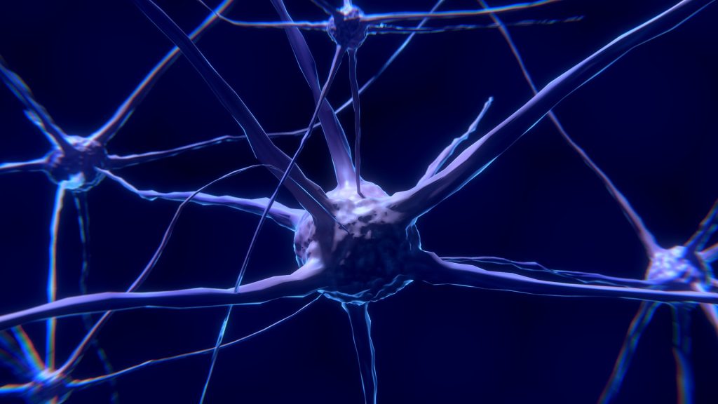
Image source: Unsplash

Researchers have found that neuron connectivity patterns within brain regions can better indicate disease progression and treatment outcomes for people with brain disorders such as epilepsy.

Many brain diseases lead to cell death and the removal of connections within the brain. A team led by Dr Marcus Kaiser from the School of Medicine at the University of Nottingham looked at epilepsy patients undergoing surgery. Their findings were published in Human Brain Mapping.

They found that changes in the local network within brain regions can predict disease progression, and also whether surgery will be successful or not.

The team found that looking at connectivity within regions of the brain, showed superior results compared to only observing fibre tract connectivity between brain regions, which is the current method. Dividing the surface of the brain into 50 000 network nodes of comparable size, each brain region could be studied as a local network with 100-500 nodes. There were distinct changes seen in these local networks in patients suffering from epileptic seizures.

Employing diffusion tensor imaging, a special measurement protocol for MRI scanners, the team of scientists showed that fibres within and between brain regions are removed for patients.

However, they found that connectivity within regions better predicted whether surgical removal of brain tissue was successful in preventing future seizures.

Dr Kaiser, Professor of Neuroinformatics at the University of Nottingham, explained: “When someone has an epileptic seizure, it ‘spreads’ through the brain. We found that local network changes occurred for regions along the main spreading pathways for seizures. Importantly, regions far away from the starting point of the seizure, for example in the opposite brain hemisphere, were involved.

“This indicates that the increased brain activity during seizures leads to changes in a wide range of brain regions. Furthermore, the longer patients suffered, the more regions showed local changes and the more severe were these changes.”

The researchers from the involved universities, along with the company Biomax, evaluated the scans of 33 temporal lobe epilepsy patients and 36 control subjects.

Project partners used the NeuroXM™ knowledge management platform to develop a knowledge model for high-resolution connectivity with more than 50 000 cortical nodes and several millions of connections and corresponding automated processing pipelines accessible through Biomax’s neuroimaging product NICARA™.

Project manager Dr Markus Butz-Ostendorf from Biomax said: “Our software can be easily employed at hospitals and can also be combined with other kinds of data from genetics or from other imaging approaches such as PET, CT, or EEG.”

Professor Yanjiang Wang, who is one of the corresponding authors, and Ms Xue Chen, both from China University of Petroleum (East China), commented: “Local connectivity was not only better in overall predictions but particularly successful in identifying patients where surgery did not lead to any improvement, identifying 95% of such cases compared to 90% when used connectivity between regions”.

Source: University of Nottingham

Why Nerves Fail to Regenerate

Image source: Pixabay

Though there are many reasons why nerves fail to regenerate, researchers from Ruhr-Universität Bochum (RUB) have made a breakthrough in their discovery a new mechanism which could lead to effective treatments.

Damage to nerve fibers in the central nervous system – brain, spinal cord, or optic nerve– often results in lifelong and severe disabilities, such as paraplegia or blindness. Though there are various known reasons why nerves fail to regenerate, treating them has not thus far not resulted in success.

Now, the RUB researchers have discovered that nerves release a protein at the injury site that attracts growing nerve fibres — and keeps them entrapped there. This prevents them from growing in the right direction to bridge the injury. Their findings are published in the journal Proceedings of the National Academy of Science (PNAS).

There are three known main causes for the inability of injured nerves of the central nervous system (CNS) to regenerate: insufficient activation of a regeneration program in injured nerve cells that stimulates the growth of fibres, so-called axons; scar formation at the injury site that is difficult for nerve fibres to penetrate; and an inhibitory effect of molecules in the nerve on regrowing axons. “Although experimental approaches have been found in recent decades to address these individual aspects by therapeutic means, even combinatorial approaches have shown only little success,” said Fischer. “So there must be other yet unknown causes for why nerve fibres in the CNS don’t regenerate.”

Using the optic nerve as a model, the research time has now shown another — quite surprising — cause for the regenerative failure in the CNS. The underlying mechanism is not based on inhibition of axon growth, as in the previously identified causes, but instead on a positive effect of a protein at the injury site on the nerve. This molecule is a so-called chemokine known as CXCL12. “The protein actually promotes the growth of axons and attracts regenerating fibers. It is, therefore, chemoattractive,” explained lead investigator Professor Dietmar Fischer. However, this chemoattraction turned out to be more hindrance than help after nerve injury in living animals.

Nerve fibres are trapped

The scientists showed that this protein is released at the nerve’s lesion site and, as a result, keeps the axons at the injured area through the chemoattractive effect. As a result, even some fibres that had already regenerated across the injury site reversed direction, growing backwards to the injury site. The regrowing fibers thus remained trapped due to CXCL12’s attractive effect.

The researchers figured out this effect when they knocked out the receptor for CXCL12 in the retinal nerve cells, rendering them blind to this protein. “Surprisingly, this led to greatly increased fibre growth in the injured optic nerves, and axons showed significantly less regrowth back to the injury site,” Dietmar Fischer points out.

New drug possibilities

The researchers then investigated where at the injury site the CXCL12 originated. They found out that about eight percent of the nerve cells in the retina produce this protein themselves, transport it along their fibers to the injury site in the optic nerve, and release it there from the severed axons. “It is still unknown why some of these nerve cells make CXCL12 and others make the receptor,” said Prof Fischer. “We don’t yet understand the physiological role of the protein, but we can see that it is a major inhibitor of neural repair.”

In further experiments, the researchers showed that knocking out CXCL12 in retinal nerve cells to prevent its release at the injury site equally improved axonal regeneration into the optic nerve. “These new findings open the opportunity to develop pharmacological approaches aimed at disrupting the interaction of CXCL12 and its receptor on the nerve fibres, to free them from their captivity at the site of injury,” concluded Prof Fischer.

His team is now investigating whether similar approaches can also promote the regeneration of axons in other areas of the injured brain or spinal cord.

Source: Ruhr-Universität Bochum

Journal information: Alexander M. Hilla, et al. CXCR4/CXCL12-mediated entrapment of axons at the injury site compromises optic nerve regeneration, in: PNAS, 2021, DOI: 10.1073/pnas.2016409118

Researchers Close in on Genetic Cure for Congenital Deafness

Researchers are a step closer in the quest to use gene therapy to enable people born deaf to hear, having uncovered a new role for a key protein.

The study, published in Molecular Biology of the Cell, focused on a large gene responsible for an inner-ear protein called otoferlin. Otoferlin mutations are linked to severe congenital hearing loss, a common type of deafness in which patients can hear almost nothing.

“For a long time otoferlin seemed to be a one-trick pony of a protein,” explained Colin Johnson, associate professor of biochemistry and biophysics in the Oregon State UniversityCollege of Science. “A lot of genes will find various things to do, but the otoferlin gene had appeared only to have one purpose and that was to encode sound in the sensory hair cells in the inner ear. Small mutations in otoferlin render people profoundly deaf.”

Because the otoferlin gene is too big as it normally is to package into a delivery vehicle for molecular therapy, Prof Johnson’s team explored the use of a shortened version.

Research led by graduate student Aayushi Manchanda showed the shortened version needed to have part of the gene known as the transmembrane domain, for a surprising reason: without it, the sensory cells matured slowly.

“That was surprising since otoferlin was known to help encode hearing information but had not been thought to be involved in sensory cell development,” Johnson said.

For years, scientists in Prof Johnson’s lab have been working with the otoferlin molecule and in 2017 they identified a shortened form of the gene that can function in the encoding of sound.

To find out if the transmembrane domain of otoferlin needed to be part of the shortened version of the gene, Manchanda shortened the transmembrane domain in zebrafish.

Zebrafish are a small freshwater species that is very popular as a research organism. They grow rapidly, from a cell to a swimming fish in about five days, and share a remarkable similarity to humans at the molecular, genetic and cellular levels due to the conservation of mammalian genes early in their evolution. Embryonic zebrafish are transparent and easily maintained, and are amenable to genetic manipulation.

“The transmembrane domain tethers otoferlin to the cell membrane and intracellular vesicles but it was not clear if this was essential and had to be included in a shortened form of otoferlin,” Manchanda said. “We found that the loss of the transmembrane domain results in the sensory hair cells producing less otoferlin as well as deficits in hair cell activity. The mutation also caused a delay in the maturation of the sensory cells, which was a surprise. Overall the results argue that the transmembrane domain must be included in any gene therapy construct.”

At the molecular level, Manchanda found that a lack of transmembrane domain led to otoferlin not properly linking the neurotransmitter-filled synaptic vesicles to the cell membrane, resulting in less neurotransmitter being released.

“Our study suggests otoferlin’s ability to tether the vesicles to the cell membrane is a key mechanistic step for neurotransmitter release during the encoding of sound,” Manchanda said.

Source: EurekaAlert!

Two-way Signalling Discovered in Certain Neurons

It was long thought that information travelled in a one-way direction, but a new study has revealed that information also travels in the opposite direction at a key synapse in the hippocampus, the brain region responsible for learning and memory. 

Now, Peter Jonas and his group at the Institute of Science and Technology Austria (IST Austria) have demonstrated that information can also travel in the opposite direction at a key synapse in the hippocampus. At the ‘mossy fibre synapse’, the post-synaptic CA3 neuron influences the firing of the post-synaptic ‘mossy fibre neuron’. Their work was published in Nature Communications.

“We have shown, for the first time, that a retrograde information flow is physiologically relevant for pre-synaptic plasticity,” said Yuji Okamoto, a postdoc in the group of Peter Jonas at IST Austria and co-first author of the paper published in Nature Communications.

In the neuronal network, the mossy fibre synapse play a key role in information storage. Synaptic transmission is plastic, meaning that a variable amount of neurotransmitter is released into the synapse. To understand the mechanism of plasticity at work in this synapse, Okamoto precisely stimulated the pre-synaptic terminal of the mossy fibre synapse in rats and at the same time recorded electrical properties at the post-synaptic neuron. “We need to know the synapse’s exact properties—with the numerical values, eg, for its conductance—to create an exact model of this synapse. With his exact measurements, Yuji managed to obtain these numbers,” added Peter Jonas, co-corresponding author with postdoc David Vandael.

Smart teacher balances student’s workload

The researchers found that, unexpectedly, the post-synaptic neuron has an influence on plasticity in the pre-synaptic neuron. Previously the assumption was that the mossy fibre was a ‘teacher synapse’, inducing firing in the post-synaptic neuron. “Instead, we find that this synapse acts like a ‘smart teacher’, who adapts the lessons when students are overloaded with information. Similarly, the pre-synaptic mossy fibre detects when the post-synaptic neuron can’t take more information: When activity increases in the post-synaptic neuron, the pre-synaptic neuron reduces the extent of plasticity,” explained Jonas.

This finding raises the question of how the post-synaptic neuron sends information about its activity status to the pre-synaptic neuron. Pharmacological evidence suggests a role for glutamate, one of the key neurotransmitters used by neurons to send signals to other cells. Glutamate is also the transmitter released from pre-synaptic mossy fibre terminals. When calcium levels increase in the post-synaptic neuron—a sign that the neuron is active—the post-synaptic neuron may release vesicles with glutamate into the synapse. The glutamate travels back to the pre-synaptic neuron, against the usual flow of neuronal information.

“This retrograde modulation of plasticity likely helps to improve information storage in the downstream hippocampal network,” said Jonas, adding: “Once again, exact measurements have shown that reality is more complex than a simplified model would suggest.”

Source: Institute of Science and Technology Austria

Journal information: David Vandael et al. Transsynaptic modulation of presynaptic short-term plasticity in hippocampal mossy fiber synapses, Nature Communications (2021). DOI: 10.1038/s41467-021-23153-5

Brain-computer Interface Lets Paralysed People Write Letters

Image by Gerd Altmann from Pixabay

Researchers have developed a new brain-computer interface (BCI) that can let paralysed people write by mentally writing letters by hand.

Working with a participant with paralysis who has sensors implanted in his brain, the team used an algorithm to identify letters in real time as he attempted to write them, putting the results on a screen.

This technology could be further developed to allow people with paralysis type rapidly without using their hands, said study coauthor Krishna Shenoy, a Howard Hughes Medical Institute Investigator at Stanford University who jointly supervised the work with Jaimie Henderson, a Stanford neurosurgeon.

By attempting handwriting, the study participant was able to ‘type’ 90 characters per minute — more than double the previous record for typing with such a brain-computer interface.

Thought-powered communication

Even if injury or disease the ability to move, the brain’s neural activity for being able to do so remains. By making use of this activity, researchers can help people with paralysis or amputations regain lost abilities.

In recent years, Shenoy’s team has decoded the neural activity associated with speech in the hopes of reproducing it. Patients with implanted sensors mentally pointed at and clicked on letters on a screen to type at about 40 characters per minute, the previous speed record for typing with a BCI.
Wanting to try something new and different, Frank Willett, a neuroscientist in Shenoy’s group, wondered if it might be possible to harness the brain signals evoked by writing by hand “We want to find new ways of letting people communicate faster,” he said. 

The team worked with a participant enrolled in a clinical trial involving BCIs. Henderson implanted two tiny sensors into the part of the brain that controls the hand and arm, making it possible for the person to, for example, move a robotic arm or a cursor on a screen by attempting to move their own paralysed arm.
The participant, who was 65 years old at the time of the research, had a spinal cord injury that left him paralysed from the neck down. A machine learning algorithm recognised the patterns his brain produced when he attempted to write each letter.

With this system, the man could copy sentences and answer questions at a rate similar to that of someone his age typing on a smartphone. The reason why this so-called “Brain-to-Text” BCI is so fast is because each letter elicits a highly distinctive activity pattern, making it relatively easy for the algorithm to distinguish one from another, Willett explained.

A new system

Shenoy’s team envisions using attempted handwriting for text entry as part of a more comprehensive system that also includes point-and-click navigation, much like that used on current smartphones, and even attempted speech decoding. “Having those two or three modes and switching between them is something we naturally do,” he said.
The team intends to next work with a participant who cannot speak, such as a person with amyotrophic lateral sclerosis, a degenerative neurological disorder leading to loss of movement and speech.

The new system could potentially help those suffering from paralysis caused by a number of conditions, Henderson added. Those include brain stem stroke, which afflicted Jean-Dominique Bauby, the author of the book The Diving Bell and the Butterfly. “He was able to write this moving and beautiful book by selecting characters painstakingly, one at a time, using eye movement,” Henderson said. “Imagine what he could have done with Frank’s handwriting interface!”

Source: Howard Hughes Medical Institute

Scientists Find How Enriched Environments Boost Brains

Image by Colin Behrens from Pixabay

A recent study in Frontiers in Molecular Neuroscience has shown how environmental enrichment ‘opens up’ chromosomes through the action of ‘master switches’.

Environmental enrichment, that is, making stimulating and interesting surroundings, is often used in zoos, laboratories, and farms to stimulate animals and increase their wellbeing.

Stimulating environments are better for mental health and cognition because they boost the growth and function of neurons and their connections, the glia cells that support and feed neurons, and blood vessels within the brain. But what are the deeper molecular mechanisms that first set in motion these large changes in neurophysiology? 

The study investigators utilised a large molecular toolbox to map how environmental enrichment leads to changes in the 3D organisation of chromosomes in neurons and glia cells of the mouse brain, which change the activation of some genes within the genome. 

They show that genes which in humans are important for cognitive mental health are particularly affected, possibly leading to new treatments.

Chromosome ‘opens up’ with enrichment

“Here we show for the first time, with large-scale data from many state-of-the-art methods, that young adolescent mice that grew up in an extra stimulating environment have highly specific ‘epigenetic’ changes—that is, molecular changes other than in DNA sequence—to the chromosomes within the cells of the brain cortex,” said corresponding author Dr Sergio Espeso-Gil from the Centre for Genomic Regulation in Barcelona, Spain.

He continued, “These increase the local ‘openness’ and ‘loopiness’ of the chromosomes, especially around DNA stretches called enhancers and insulators, which then fine-tune more ‘downstream’ genes. This happens not only in neurons but also in the supportive glia cells, too often ignored in studies about learning.”

The team raised mice for the first month after birth in social groups inside housing with Lego blocks, ladders, balls, and tunnels that were frequently changed and moved around. As a control, other mice were raised in smaller groups inside standard housing. The researchers then used a variety of tools to pick up molecular changes in neurons and glia cells within the brain cortex. These included alterations in the 3D structure of chromosomes, particularly the local “chromatin accessibility” (openness) and “chromatin interactions” (where distant genes are brought together through loops, to coordinate activity). Chromatins are the proteins which make up chromosomes, carrying DNA and the proteins to package them.

Epigenetic ‘master’ switches

They show that two ‘master’ switches operational after environmental enrichment increase chromatin interactions and another increases chromatin availability, important for the pyramidal neurons involved in cognition. A third works on a key chromosomal protein histone H3, activating nearby genes as a result.

These switches mainly occur around genomic regions that contain enhancers, regulatory DNA that (when bound to proteins called transcription factors) can activate neighboring genes. Also affected were genomic regions with insulators, regulatory DNA that can override the gene-activating effect of neighboring enhancers.

The team concluded that growing up in an enriched environment causes highly local and specific epigenetic changes in neurons and glia cells. These then mostly increase the activity of a few genes within the genome.

Mental health in humans

“Our results show that many of the genes involved are known to play a role in the growth and differentiation of neurons, the development of blood vessels, the formation and patterning of new synaptic connections on neurons, and molecular pathways implicated in memory and learning in mice,” said Dr Espeso-Gil.

“And when we look for parallel regions in the human genome, we find many regions that are statistically associated with differences in complex traits such as insomnia, schizophrenia, and Alzheimer’s in humans, which means that our study could inform future research on these disorders. This points to the potential of environmental enrichment in therapies for mental health. Our research could also help to guide future research on chromatin interactions and the poorly known importance of glial cells for cognitive mental health.”

Source: Medical Xpress

Journal information: Sergio Espeso-Gil et al, Environmental Enrichment Induces Epigenomic and Genome Organization Changes Relevant for Cognition, Frontiers in Molecular Neuroscience (2021). DOI: 10.3389/fnmol.2021.664912

Study Explores the Circadian Rhythm Control Centre

Woman sleeping with an alarm clock on bedside. Photo by cottonbro from Pexels

Researchers in Japan have offered new insights into how the brain’s circadian rhythm control centre regulates behaviour.

Circadian rhythms are a force in the background that shapes many human behaviours such feeling tired and falling asleep, as well as influencing our health. Michihiro Mieda and his team at Kanazawa University in Japan are researching just how the brain’s circadian rhythm control centre regulates behaviour.

The control centre, known as the superchiasmatic nucleus, or SCN, contains many types of neurons that transmit signals using the molecule GABA, but little is known about how each type contributes to our bodily rhythms. In this most recent study, the researchers focused on GABA neurons that produce arginine vasopressin, a hormone that regulates kidney function and blood pressure in the body, and which the team recently showed is also involved in the regulation of the interval of rhythms produced by the SCN.

To examine the function of these neurons separate to all others, the researchers first deleted a gene in mice which was needed for GABA signaling between neurons, but only in vasopressin-producing SCN neurons. “We removed a gene that codes for a protein that allows GABA to be packaged before it is sent to other neurons,” explained Mieda. “Without packaging, none of the vasopressin neurons could send out any GABA signals.”

Thus, these neurons could not use GABA to communicate with the rest of the SCN anymore. The mice showed longer periods of activity, beginning activity earlier and ending activity later than control mice, a simple enough result. It might seem that losing the packaging gene in the neurons disrupted the molecular clock signal but the result was not so simple. Closer examination deepened the mystery as the molecular clock seemed to progress unhindered.
Using calcium imaging, the researchers examined the clock rhythms within the vasopressin neurons. They found that while the rhythm of activity matched the timing of behaviour in control mice, this relationship was disturbed in the mice with missing GABA transmission in the vasopressin neurons. The rhythm of SCN output, ie SCN neuronal electrical activity, in the modified mice had the same irregular rhythm as their behaviour.

“Our study shows that GABA signaling from vasopressin neurons in the suprachiasmatic nucleus help fix behavioral timing within the constraints of the molecular clock,” concluded Mieda.

Source: News-Medical.Net

Journal reference: Maejima, T., et al. (2021) GABA from vasopressin neurons regulates the time at which suprachiasmatic nucleus molecular clocks enable circadian behavior. PNAS. doi.org/10.1073/pnas.2010168118.

Scientists Crack Neuron Information Storage Code

A team of scientists from the UK and Australia have discovered that single neurons can store electrical patterns, similar to memories. This represents a breakthrough towards solving how neural systems are able to process and store information.

By comparing predictions from mathematical modeling to lab-based experiments with mammalian neurons, they were able to determine how different parameters, such as how long it takes for neuronal signals to be processed and how sensitive a cell is to external signals, affect how neural systems encode information.

The research team found that a single neuron is able to select between different patterns, dependent on the properties of each individual stimulus, for example slight differences in stimulation timing resulted in the emergence of no electrical activity spikes, single spikes per delay or two spikes per delay,

By opening up new avenues into research on the encoding of information in the brain and how this relates to memory formation, the study could also allow new insights into the causes and treatments of mental health conditions such as dementia.

“This work highlights how mathematical analysis and wet-lab experiments can be closely integrated to shed new light on fundamental problems in neuroscience,” said Dr Wedgwood. “That the theoretical predictions were so readily confirmed in experiments gives us great confidence in the mathematical approach as a tool for understanding how individual cells store patterns of activity. In the long run, we hope that this is the first step to a better understanding of memory formation in neural networks.”

Professor Krauskopf from the University of Auckland remarked, “The research shows that a living neuron coupled to itself is able to sustain different patterns in response to a stimulus. This is an exciting first step towards understanding how groups of neurons are able to respond to external stimuli in a precise temporal manner.”

“Communication between neurons occurs over large distances. The communication delay associated with this plays an important role in shaping the overall response of a network. This insight is crucial to how neural systems encode memories, which is one of the most fundamental questions in neuroscience,” added Professor Tsaneva from the University of Exeter’s Living Systems Institute.

Source: Medical Xpress

Journal information: Kyle C. A. Wedgwood et al, Robust spike timing in an excitable cell with delayed feedback, Journal of The Royal Society Interface (2021). dx.doi.org/10.1098/rsif.2021.0029

Eye Pressure in Glaucoma not the Whole Story

The findings of a new study in rats show that a chemical known to protect nerve cells also slows glaucoma, the leading cause of irreversible blindness.

According to the National Glaucoma Foundation, in the US, over 3 million have glaucoma, with only half being aware of the fact and more than 120 000 are blind from the disease. The World Health Organization estimates that, worldwide, over 60 million individuals suffer from glaucoma.  

Led by researchers at NYU Grossman School of Medicine, the study centred on the watery fluid inside the eye on which its function depends. In patients with glaucoma, a buildup of fluid pressure wears down cells in the eyes and the nerves connecting them to the brain.

Previous research that despite eye pressure having been controlled, the condition progressively worsened. The relationship between pressure buildup and impaired vision remains poorly understood

The new study showed that when rats ingested the compound citicoline, optic nerve signals between the brain and eye were almost fully restored. Citicoline is a major source of choline, a building block in the membranes that line nerve cells and enhance nerve cell communication. It is produced in the brain but also commercially produced.

The study confirmed that increased eye pressure levels contributes to nerve damage in glaucoma, but  it also showed that citicoline reduced vision loss in rats without reducing pressure levels.

“Our study suggests that citicoline protects against glaucoma through a mechanism different from that of standard treatments that reduce fluid pressure,” said senior author Kevin Chan, PhD, an assistant professor in the Department of Ophthalmology at NYU Langone Health. “Since glaucoma interrupts the connection between the brain and eye, we hope to strengthen it with new types of therapies.”

The findings are helping scientists better understand how glaucoma works and add to past evidence that citicoline may counter the disease, said Chan, also the director of the Neuroimaging and Visual Science Laboratory at NYU Langone. It is known that humans and rodents with glaucoma have lower than normal levels of choline in the brain, but until now, Prof Chan says, there’s been little concrete evidence of the effectiveness of choline supplements as a therapy for glaucoma or why choline occurs in lower levels in glaucoma patients.

Prof Chan and his team tested whether increasing levels of that chemical would slow or even stop the degradation of the optic nerve and other regions of the brain involved in vision. Using a comprehensive study of the eye-brain connection in glaucoma, his team found that giving rats oral doses of citicoline over a three-week period protected nerve tissues and reduced vision loss sustainably even after the treatment stopped for another three weeks.

To simulate glaucoma, the researchers used a clear gel in rats to build up eye pressure mildly without otherwise blocking their vision. Then, the team used MRI imagery to measure the structural integrity and the amount of functional and physiological activity along the visual pathway. To test the clarity of vision of each eye, the researchers tracked the rodents’ visual behaviour .

It was found that for rats with mildly elevated eye pressure, the tissues that connect the eye and brain, including the optic nerve, degraded for up to five weeks after the injury. Nerve structure breakdown in the citicoline-treated rodents slowed by up to 74%, which the researchers said indicates that the chemical had protective effects on nerve cells.

However, more research is necessary before citicoline supplements to treat glaucoma in humans, as commercial drugs have yet to be proven fully effective in clinical trials. The researchers are planning next to look into how choline protects the eye and why it is depleted in glaucoma patients.

Source: Medical Xpress

Journal information: Yolandi van der Merwe et al, Citicoline Modulates Glaucomatous Neurodegeneration Through Intraocular Pressure-Independent Control, Neurotherapeutics (2021). DOI: 10.1007/s13311-021-01033-6

Impairment Lasts up to 10 Hours After Cannabis

A comprehensive analysis of 80 scientific studies has identified a ‘window of impairment’ of between three and 10 hours caused by moderate to high doses of tetrahydrocannabinol (THC), the cannabis component that causes intoxication. According to the researchers, these results have consequences for drug-driving laws around the world.

How long the impairment lasts depends on the THC dose, whether it is taken orally or inhaled, on the usage habits of the cannabis user and the demands of the task. The psychoactive THC component of cannabis has potential medical applications in treating nausea, sleep apnoea, fibromyalgia and chronic pain, though these applications are controversial and currently difficult to study due to legal issues, though off-label use is common. 
Previous research by Dr Arkell and colleagues has shown that cannabidiol (CBD), one of the medically active components of cannabis, does not cause impairment in driving. CBD has analgesic and anti-inflammatory actions, as well as anxiolytic, antiemetic, antipsychotic, and neuroprotective antioxidant properties

Medical and non-medical legal cannabis use is on the rise worldwide.
THC causes acute impairment in driving and cognitive performance, but there is uncertainty among users about the duration of this impairment and when they can start tasks such as driving after consuming cannabis.
“Our analysis indicates that impairment may last up to 10 hours if high doses of THC are consumed orally,”  said lead author Dr Danielle McCartney, Lambert Initiative for Cannabinoid Therapeutics at the University of Sydney. “A more typical duration of impairment, however, is four hours, when lower doses of THC are consumed via smoking or vaporization and simpler tasks are undertaken (eg, those using cognitive skills such as reaction time, sustained attention and working memory). This impairment may extend up to six or seven hours if higher doses of THC are inhaled and complex tasks, such as driving, are assessed.”

A moderate THC dose is considered about 10 milligrams in this study, but could be higher for a regular user, said the researchers.

Co-author Dr Thomas Arkell, also from the Lambert Initiative, said: “We found that impairment is much more predictable in occasional cannabis users than regular cannabis users. Heavy users show significant tolerance to the effects of cannabis on driving and cognitive function, while typically displaying some impairment.”

Regular cannabis users might consume more to get the same effect, resulting in equivalent impairment, the authors noted.

In the case of oral use as in medical cannabis drops, tablets etc, the impairment takes longer to manifest and has a longer duration than the inhalation route.

The findings have implications for so-called drug-driving laws, the researchers said.

Professor Iain McGregor, Academic Director of the Lambert Initiative, said: “THC can be detected in the body weeks after cannabis consumption while it is clear that impairment lasts for a much shorter period of time. Our legal frameworks probably need to catch up with that and, as with alcohol, focus on the interval when users are more of a risk to themselves and others. Prosecution solely on the basis of the presence of THC in blood or saliva is manifestly unjust.

“Laws should be about safety on the roads, not arbitrary punishment. Given that cannabis is legal in an increasing number of jurisdictions, we need an evidence-based approach to drug-driving laws,” Prof McGregor said.

Source: News-Medical.Net

Journal information: McCartney, D., et al. (2021) Determining the magnitude and duration of acute Δ9-tetrahydrocannabinol (Δ9-THC)-induced driving and cognitive impairment: A systematic and meta-analytic review. Neuroscience & Biobehavioral Reviews. doi.org/10.1016/j.neubiorev.2021.01.003.